Copyright
©The Author(s) 2015.
World J Gastrointest Endosc. Nov 25, 2015; 7(17): 1257-1261
Published online Nov 25, 2015. doi: 10.4253/wjge.v7.i17.1257
Published online Nov 25, 2015. doi: 10.4253/wjge.v7.i17.1257
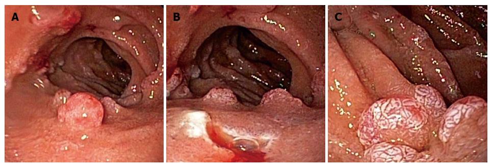
Figure 1 Duodenal polyposis under esophagogastroduodenoscopy.
A 6 mm, sessile polyp was seen [prior to removal (A); immediately after removal (B)] in the mid duodenal bulb. A separate 8 mm polyp was seen along the lateral aspect of the second part of the duodenum (C).
- Citation: Gurung A, Jaffe PE, Zhang X. Duodenal polyposis secondary to portal hypertensive duodenopathy. World J Gastrointest Endosc 2015; 7(17): 1257-1261
- URL: https://www.wjgnet.com/1948-5190/full/v7/i17/1257.htm
- DOI: https://dx.doi.org/10.4253/wjge.v7.i17.1257


