Copyright
©The Author(s) 2015.
World J Gastrointest Endosc. Nov 10, 2015; 7(16): 1208-1215
Published online Nov 10, 2015. doi: 10.4253/wjge.v7.i16.1208
Published online Nov 10, 2015. doi: 10.4253/wjge.v7.i16.1208
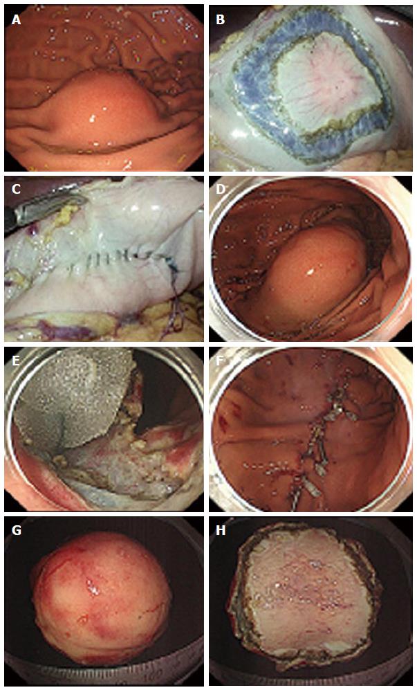
Figure 5 Procedures of non-exposed endoscopic wall-inversion surgery.
A: Protruding submucosal lesion is seen at the greater curvature of the middle gastric body; B: Circumferential seromuscular incision is made outside the serosal markings after endoscopic submucosal injection; C: Lesion is inverted with a surgical sponge used as a spacer; D: Massive protrusion of the inverted tissue; E: Surgical sponge as a spacer and a suturing line during endoscopic mucosal incision; F: Mucosal clipping after the resection; G: Resected specimen: Mucosal side; H: Resected specimen: Serosal side.
- Citation: Maehata T, Goto O, Takeuchi H, Kitagawa Y, Yahagi N. Cutting edge of endoscopic full-thickness resection for gastric tumor. World J Gastrointest Endosc 2015; 7(16): 1208-1215
- URL: https://www.wjgnet.com/1948-5190/full/v7/i16/1208.htm
- DOI: https://dx.doi.org/10.4253/wjge.v7.i16.1208


