Copyright
©The Author(s) 2015.
World J Gastrointest Endosc. Oct 25, 2015; 7(15): 1170-1180
Published online Oct 25, 2015. doi: 10.4253/wjge.v7.i15.1170
Published online Oct 25, 2015. doi: 10.4253/wjge.v7.i15.1170
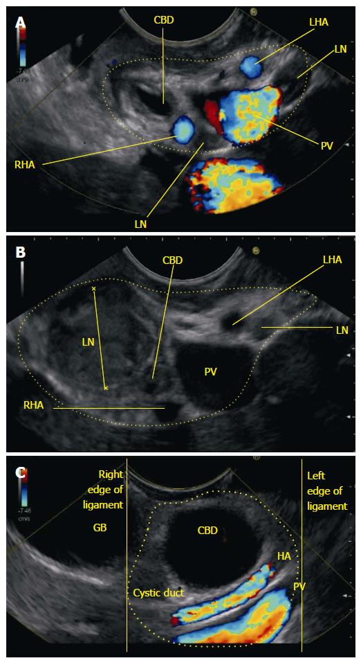
Figure 16 Hepatoduodenal ligament.
A: The anticlockwise rotation takes the probe towards the hilum of liver the bile duct is demonstrated in a transverse axis. The HDL contains the structures of portal triad; B: Further anticlockwise rotation shows an abnormal lymph node within the hepatoduodenal ligament which is causing obstruction of CBD; C: On further anticlockwise rotation the probe moves towards the hilum of liver and the dilated bile duct is demonstrated in a transverse axis along with cystic duct and gall bladder. The portal vein and hepatic artery are demonstrated in long axis. All these structures shown lie in the hepatoduodenal ligament near the hilum except the gallbladder. CBD: Common bile duct; CHD: Common hepatic duct; RHA: Right hepatic artery; LHA: Left hepatic artery; LN: Lymph node; PV: Portal vein; HDL: Hepatoduodenal ligament.
- Citation: Sharma M, Pathak A, Shoukat A, Rameshbabu CS, Ajmera A, Wani ZA, Rai P. Imaging of common bile duct by linear endoscopic ultrasound. World J Gastrointest Endosc 2015; 7(15): 1170-1180
- URL: https://www.wjgnet.com/1948-5190/full/v7/i15/1170.htm
- DOI: https://dx.doi.org/10.4253/wjge.v7.i15.1170


