Copyright
©The Author(s) 2015.
World J Gastrointest Endosc. Oct 25, 2015; 7(15): 1170-1180
Published online Oct 25, 2015. doi: 10.4253/wjge.v7.i15.1170
Published online Oct 25, 2015. doi: 10.4253/wjge.v7.i15.1170
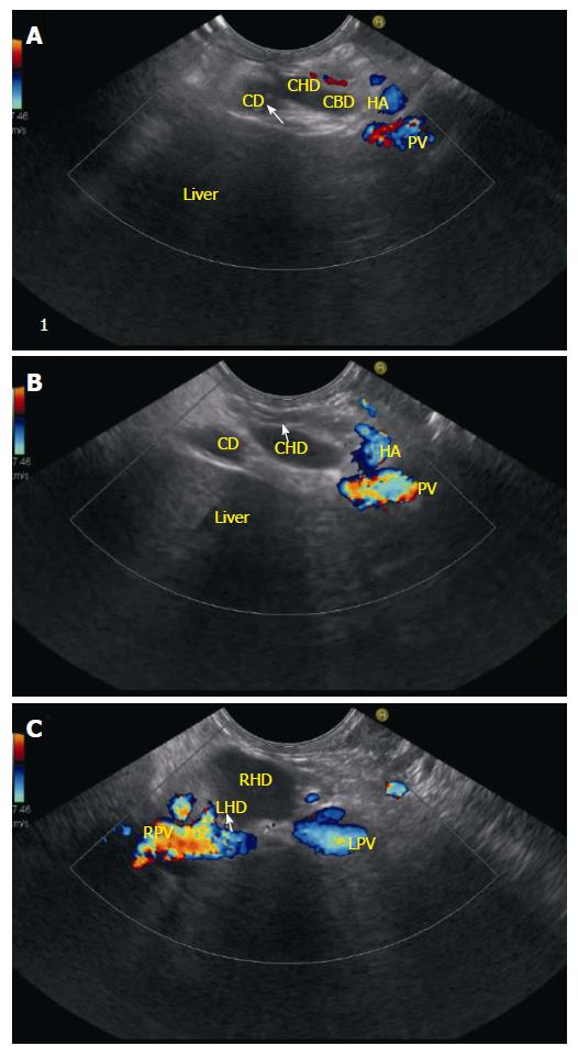
Figure 15 Cystic duct and common hepatic duct imaging.
A and B: The cystic duct terminates at the right wall of the common hepatic duct in 85 to 90% of cases. In this case the CD is seen joining the right aspect of CHD; C: When the CHD is followed up it divides into right and left hepatic duct and this bifurcation generally lies in front of the right branch of the portal vein. As the echoendoscope is rotated counter clockwise the portal vein is followed up to its bifurcation and the RPV is seen on the right side of the screen. CHD: Common hepatic duct; CD: Cystic duct; CBD: Common bile duct; PV: Portal vein; HA: Hepatic artery; RHD: Right hepatic duct; LHD: Left Hepatic duct; LPV: Left portal vein; RPV: Right portal vein.
- Citation: Sharma M, Pathak A, Shoukat A, Rameshbabu CS, Ajmera A, Wani ZA, Rai P. Imaging of common bile duct by linear endoscopic ultrasound. World J Gastrointest Endosc 2015; 7(15): 1170-1180
- URL: https://www.wjgnet.com/1948-5190/full/v7/i15/1170.htm
- DOI: https://dx.doi.org/10.4253/wjge.v7.i15.1170


