Copyright
©The Author(s) 2015.
World J Gastrointest Endosc. Oct 25, 2015; 7(15): 1170-1180
Published online Oct 25, 2015. doi: 10.4253/wjge.v7.i15.1170
Published online Oct 25, 2015. doi: 10.4253/wjge.v7.i15.1170
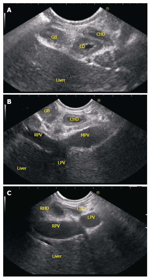
Figure 14 Hilum imaging from D2.
A: Anticlockwise rotation from the 2nd part of duodenum traces the CBD towards the hilum. The cystic duct is seen taking origin from the aspect away from transducer and the gall bladder is visualized; B: When imaging is done from below upwards the imaging shows the CHD going towards the right portal vein; C: Further anticlockwise rotation towards hilum can show the left and right hepatic duct. The division of CHD into RHD and LHD occurs in front of right branch of portal vein. CBD: Common bile duct; CHD: Common hepatic duct; RHD: Right hepatic duct; LHD: Left hepatic duct; LPV: Left portal vein; RPV: Right portal vein.
- Citation: Sharma M, Pathak A, Shoukat A, Rameshbabu CS, Ajmera A, Wani ZA, Rai P. Imaging of common bile duct by linear endoscopic ultrasound. World J Gastrointest Endosc 2015; 7(15): 1170-1180
- URL: https://www.wjgnet.com/1948-5190/full/v7/i15/1170.htm
- DOI: https://dx.doi.org/10.4253/wjge.v7.i15.1170


