Copyright
©The Author(s) 2015.
World J Gastrointest Endosc. Oct 25, 2015; 7(15): 1170-1180
Published online Oct 25, 2015. doi: 10.4253/wjge.v7.i15.1170
Published online Oct 25, 2015. doi: 10.4253/wjge.v7.i15.1170
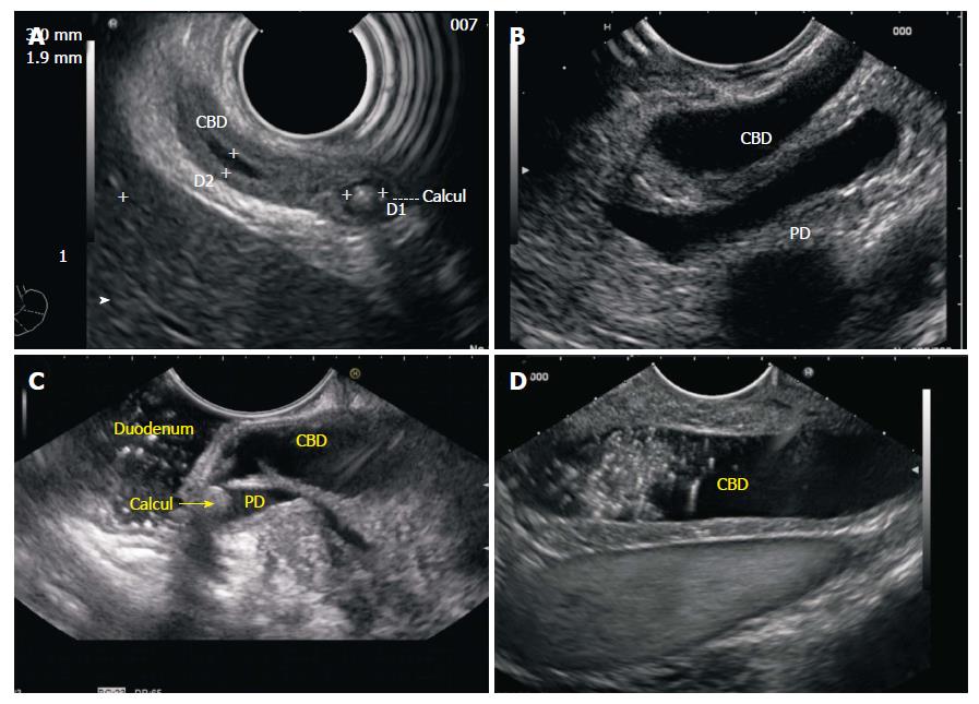
Figure 12 Imaging from 2nd part of duodenum.
A: The intrapapillary part of CBD and the lower 1/3 of CBD is sometimes best visualized with the radial EUS scope as they provide a long axis of imaging of the entire bile duct in a long axis; B: However good view of CBD in a long axis can be also obtained by linear EUS scope. This image shows the dilated CBD and PD in a long axis in a case of periampullary carcinoma; C: The distended CBD may not provide room for good visualization as it comes very close to probe in a pathology involving papilla. This figure shows good view of CBD after instillation of 100 mL water which provides good coupling and also provides adequate focal distance. The stone is impacted in the common channel where it is also obstructing and dilating the pancreatic duct; D: The dilated CBD with sludge is seen from 2nd part of duodenum. On the far side of screen the IVC is also seen beyond the IVC. CHD: Common hepatic duct; EUS: Endoscopic ultrasound; IVC: Inferior venacava; CBD: Common bile duct; PD: Pancreatic duct.
- Citation: Sharma M, Pathak A, Shoukat A, Rameshbabu CS, Ajmera A, Wani ZA, Rai P. Imaging of common bile duct by linear endoscopic ultrasound. World J Gastrointest Endosc 2015; 7(15): 1170-1180
- URL: https://www.wjgnet.com/1948-5190/full/v7/i15/1170.htm
- DOI: https://dx.doi.org/10.4253/wjge.v7.i15.1170


