Copyright
©The Author(s) 2015.
World J Gastrointest Endosc. Oct 25, 2015; 7(15): 1170-1180
Published online Oct 25, 2015. doi: 10.4253/wjge.v7.i15.1170
Published online Oct 25, 2015. doi: 10.4253/wjge.v7.i15.1170
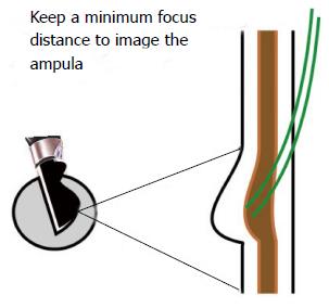
Figure 10 Imaging of Common hepatic duct at papilla can be done after apposition of transducer with the papilla, which is the main endoscopic landmark.
It is appreciated as a thickening of the duodenal wall and a rounded 5-layered structure. Good views of papilla require three things: (1) transducer perpendicular to papilla; (2) good water coupling; (3) and motionless duodenum). If a balloon is used only a small amount of water should be filled in balloon to avoid smashing the delicate papilla. The imaging of papilla after instillation of about 50 to 100 mL water keeps the transducer away from papilla, increases the focal distance of imaging of transducer from papilla and places the papilla as well as lower 1/3 of CBD in the optimum focal distance of imaging (usually about 1 cm). CHD: Common hepatic duct.
- Citation: Sharma M, Pathak A, Shoukat A, Rameshbabu CS, Ajmera A, Wani ZA, Rai P. Imaging of common bile duct by linear endoscopic ultrasound. World J Gastrointest Endosc 2015; 7(15): 1170-1180
- URL: https://www.wjgnet.com/1948-5190/full/v7/i15/1170.htm
- DOI: https://dx.doi.org/10.4253/wjge.v7.i15.1170


