Copyright
©The Author(s) 2015.
World J Gastrointest Endosc. Oct 25, 2015; 7(15): 1170-1180
Published online Oct 25, 2015. doi: 10.4253/wjge.v7.i15.1170
Published online Oct 25, 2015. doi: 10.4253/wjge.v7.i15.1170
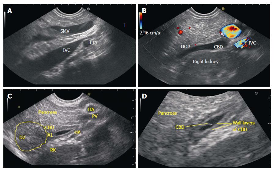
Figure 6 Lower Common bile duct imaging from D2-D3.
A: In the hilum of liver the CBD lies anterior to the portal vein and both CBD and portal vein are positioned anterior to inferior vena cava. As the CBD is followed down towards ampulla the IVC remains goes posterior to head of pancreas whereas the SMV (followed down as a continuation of portal vein) comes to lie anterior to posterior part of head of pancreas. The CBD occupies the area of posterior part of head of pancreas between the SMV and IVC. This figure shows the typical appearance of SMV lying in front of IVC from stomach. If it is difficult to trace the course of CBD, the IVC, portal vein or superior mesenteric vein can be followed as a vascular home bases for tracing of CBD; B: In this figure the CBD is identified in posterior part of head of pancreas with slight anticlockwise rotation after visualizing the typical appearance of SMV lying in front of IVC; C: Once the Lower 1/3 of CBD is located it can be followed down towards the intrapancreatic part of CBD and zooming can help in imaging of papilla as well as 2nd part of duodenum; D: With selective zooming of bile duct the individual layers of bile duct can be identified. SMV: Superior mesenteric vein; CBD: Common bile duct; IVC: Inferior venacava; HOP: Head of pancreas; BOP: Body of pancreas; PV: Portal vein.
- Citation: Sharma M, Pathak A, Shoukat A, Rameshbabu CS, Ajmera A, Wani ZA, Rai P. Imaging of common bile duct by linear endoscopic ultrasound. World J Gastrointest Endosc 2015; 7(15): 1170-1180
- URL: https://www.wjgnet.com/1948-5190/full/v7/i15/1170.htm
- DOI: https://dx.doi.org/10.4253/wjge.v7.i15.1170


