Copyright
©The Author(s) 2015.
World J Gastrointest Endosc. Oct 25, 2015; 7(15): 1170-1180
Published online Oct 25, 2015. doi: 10.4253/wjge.v7.i15.1170
Published online Oct 25, 2015. doi: 10.4253/wjge.v7.i15.1170
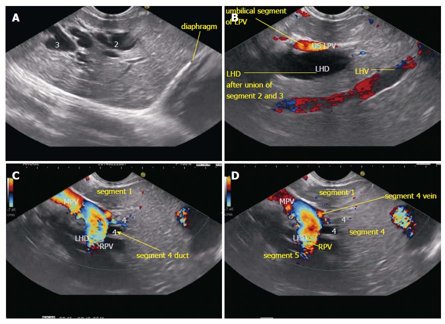
Figure 2 Segmental ducts as seen on endoscopic ultrasound.
A: The dilated ducts of segment 2 and 3 ducts are seen in an open position to left; B: On clockwise rotation the segment 2 and 3 ducts fuse together in front of umbilical part of left portal vein. The left hepatic vein is also identified going from 2 o’clock position to 7 o’clock position; C: On further clockwise rotation the fused part of segment 2 and 3 ducts is joined by segment 4 duct from the cranial aspect (arrow) in front of the transverse segment of left portal vein; D: On further clockwise rotation the right portal vein is seen joining the left portal vein and the liver segment lying below the plane of right portal vein belongs to segment 5. RPV: Right portal vein; LPV: Left portal vein; LHD: Left hepatic duct; LHV: Left hepatic vein.
- Citation: Sharma M, Pathak A, Shoukat A, Rameshbabu CS, Ajmera A, Wani ZA, Rai P. Imaging of common bile duct by linear endoscopic ultrasound. World J Gastrointest Endosc 2015; 7(15): 1170-1180
- URL: https://www.wjgnet.com/1948-5190/full/v7/i15/1170.htm
- DOI: https://dx.doi.org/10.4253/wjge.v7.i15.1170


