Copyright
©The Author(s) 2015.
World J Gastrointest Endosc. Oct 10, 2015; 7(14): 1114-1128
Published online Oct 10, 2015. doi: 10.4253/wjge.v7.i14.1114
Published online Oct 10, 2015. doi: 10.4253/wjge.v7.i14.1114
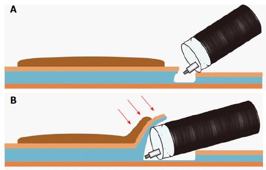
Figure 3 Schema of the mucosal flap.
A: After injecting a solution in the submucosal layer, mucosal incision and deeper cut are made; B: Continuing to dissect the submucosal layer allows the creation of the “mucosal flap” (Red arrows point to the “mucosal flap”). Inserting the distal attachment under the mucosal flap provides good counter-traction to the submucosal layers and allows good visualization of the operative field. Therefore, completion of the mucosal flap facilitates subsequent submucosal dissection.
- Citation: Yamamoto K, Michida T, Nishida T, Hayashi S, Naito M, Ito T. Colorectal endoscopic submucosal dissection: Recent technical advances for safe and successful procedures. World J Gastrointest Endosc 2015; 7(14): 1114-1128
- URL: https://www.wjgnet.com/1948-5190/full/v7/i14/1114.htm
- DOI: https://dx.doi.org/10.4253/wjge.v7.i14.1114


