Copyright
©The Author(s) 2015.
World J Gastrointest Endosc. Sep 25, 2015; 7(13): 1107-1113
Published online Sep 25, 2015. doi: 10.4253/wjge.v7.i13.1107
Published online Sep 25, 2015. doi: 10.4253/wjge.v7.i13.1107
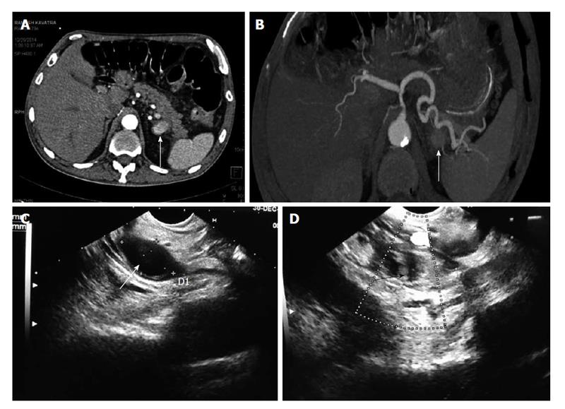
Figure 6 Chronic calcific pancreatitis with upper gastrointestinal bleed.
A: Axial contrast enhanced; B: Axial MIP (3-D reformatted) CT images; C and D: Endoscopic ultrasound images. There is a pseudoaneurysm sac (arrow, A) in relation to distal body of the pancreas, in close relation to the splenic artery (arrow, B) and confirmed by endoscopic ultrasound (arrow, C) and subsequently managed by instillation of thrombin under EUS guidance with complete thrombosis of the pseudoaneurysm was achieved as revealed by the transformation of the previously anechoic lesion to an echogenic sac (arrow, D). CT: Computed tomography; MIP: Maximum intensity projection; EUS: Endoscopic ultrasound.
- Citation: Gamanagatti S, Thingujam U, Garg P, Nongthombam S, Dash NR. Endoscopic ultrasound guided thrombin injection of angiographically occult pancreatitis associated visceral artery pseudoaneurysms: Case series. World J Gastrointest Endosc 2015; 7(13): 1107-1113
- URL: https://www.wjgnet.com/1948-5190/full/v7/i13/1107.htm
- DOI: https://dx.doi.org/10.4253/wjge.v7.i13.1107


