Copyright
©The Author(s) 2015.
World J Gastrointest Endosc. Sep 25, 2015; 7(13): 1107-1113
Published online Sep 25, 2015. doi: 10.4253/wjge.v7.i13.1107
Published online Sep 25, 2015. doi: 10.4253/wjge.v7.i13.1107
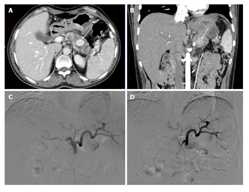
Figure 4 Pancreatitis related pseudoaneurysm (case 2).
A: Axial contrast enhanced CT; B: Coronal maximum intensity projection CT images; C and D: Digital substraction Images. In the background of pancreatitis there is a pseudoaneurysm in the distal body of the pancreas (arrow, A and B) which was not revealed on either left gastric artery (C) or splenic artery (D) angiograms. CT: Computed tomography.
- Citation: Gamanagatti S, Thingujam U, Garg P, Nongthombam S, Dash NR. Endoscopic ultrasound guided thrombin injection of angiographically occult pancreatitis associated visceral artery pseudoaneurysms: Case series. World J Gastrointest Endosc 2015; 7(13): 1107-1113
- URL: https://www.wjgnet.com/1948-5190/full/v7/i13/1107.htm
- DOI: https://dx.doi.org/10.4253/wjge.v7.i13.1107


