Copyright
©The Author(s) 2015.
World J Gastrointest Endosc. Sep 25, 2015; 7(13): 1107-1113
Published online Sep 25, 2015. doi: 10.4253/wjge.v7.i13.1107
Published online Sep 25, 2015. doi: 10.4253/wjge.v7.i13.1107
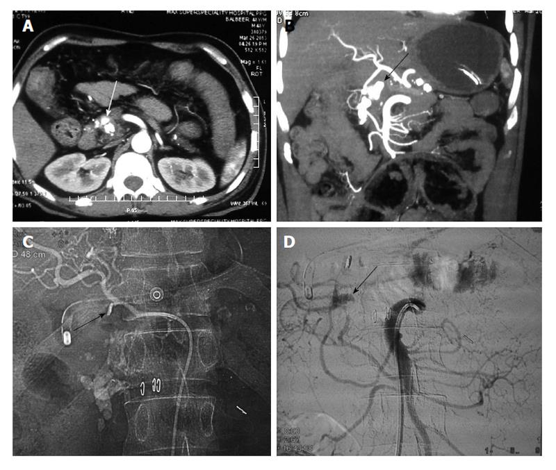
Figure 1 Chronic calcific pancreatitis with pseudoaneurysm of gastroduodenal artery (case 1).
A: Axial Contrast enhanced; B: Coronal MIP (3-D reformatted) CT images; C: Angiographic spot image; D: Superior mesenteric DSA image. There is a pseudoaneurysm (arrow, A and B) arising from the gastroduodenal artery, which is filling from pancreatic arcade (Inferior pancreatic branches from SMA, D), because of metallic clip placed surgically at the origin of gastroduodenal artery origin (arrow, C). CT: Computed tomography; MIP: Maximum intensity projection; DSA: Digital substraction angiography; SMA: Superior mesenteric artery.
- Citation: Gamanagatti S, Thingujam U, Garg P, Nongthombam S, Dash NR. Endoscopic ultrasound guided thrombin injection of angiographically occult pancreatitis associated visceral artery pseudoaneurysms: Case series. World J Gastrointest Endosc 2015; 7(13): 1107-1113
- URL: https://www.wjgnet.com/1948-5190/full/v7/i13/1107.htm
- DOI: https://dx.doi.org/10.4253/wjge.v7.i13.1107


