Copyright
©2014 Baishideng Publishing Group Inc.
World J Gastrointest Endosc. Aug 16, 2014; 6(8): 345-351
Published online Aug 16, 2014. doi: 10.4253/wjge.v6.i8.345
Published online Aug 16, 2014. doi: 10.4253/wjge.v6.i8.345
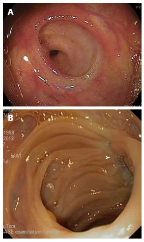
Figure 4 Endoscopic view of bilioenteric anastomosis.
A: Endoscopic view of a normal end-to-side bilioenteric anastomosis; B: Endoscopic view of a stenosis at the level of the bilioenteric anastomosis. Only scar tissue (white arrow) indicates the location of the anastomosis without visible opening.
- Citation: Moreels TG. Endoscopic retrograde cholangiopancreatography in patients with altered anatomy: How to deal with the challenges? World J Gastrointest Endosc 2014; 6(8): 345-351
- URL: https://www.wjgnet.com/1948-5190/full/v6/i8/345.htm
- DOI: https://dx.doi.org/10.4253/wjge.v6.i8.345


