Copyright
©2014 Baishideng Publishing Group Inc.
World J Gastrointest Endosc. Jul 16, 2014; 6(7): 328-333
Published online Jul 16, 2014. doi: 10.4253/wjge.v6.i7.328
Published online Jul 16, 2014. doi: 10.4253/wjge.v6.i7.328
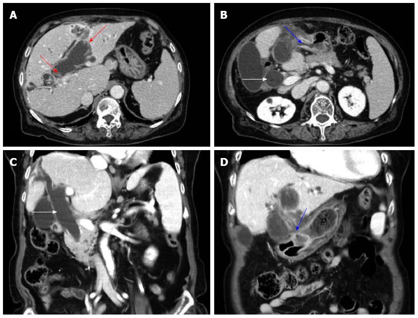
Figure 1 Computed tomography of the abdomen showed markedly dilated common bile duct (white arrows) and left intrahepatic duct with left intrahepatic duct penetrating into the antrum of stomach and fistula formation (blue arrows) and papillary projections along the dilated bile duct (red arrows) and no definite visible mass in the left intrahepatic duct (A-D).
- Citation: Hong MY, Yu DW, Hong SG. Intraductal papillary mucinous neoplasm of the bile duct with gastric and duodenal fistulas. World J Gastrointest Endosc 2014; 6(7): 328-333
- URL: https://www.wjgnet.com/1948-5190/full/v6/i7/328.htm
- DOI: https://dx.doi.org/10.4253/wjge.v6.i7.328


