Copyright
©2014 Baishideng Publishing Group Inc.
World J Gastrointest Endosc. Jul 16, 2014; 6(7): 324-327
Published online Jul 16, 2014. doi: 10.4253/wjge.v6.i7.324
Published online Jul 16, 2014. doi: 10.4253/wjge.v6.i7.324
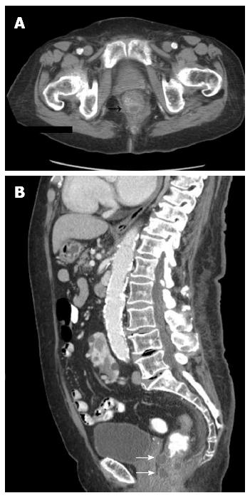
Figure 2 Computed tomography scan.
A: Computed tomography scan axial view showing mural thickening with multiple rounded hypodense foci within the posterior rectal wall; B: Computed tomography scan sagittal view showing multiple rounded foci within the anterior and posterior rectal wall (white arrows).
- Citation: Papafragkakis H, Changela K, Bhatia T, Ona MA, Malieckal A, Paleti V, Fuksbrumer MS, Anand S. Endoscopic and imaging appearance after injection of an ano-rectal bulking agent. World J Gastrointest Endosc 2014; 6(7): 324-327
- URL: https://www.wjgnet.com/1948-5190/full/v6/i7/324.htm
- DOI: https://dx.doi.org/10.4253/wjge.v6.i7.324


