Copyright
©2014 Baishideng Publishing Group Co.
World J Gastrointest Endosc. Apr 16, 2014; 6(4): 128-136
Published online Apr 16, 2014. doi: 10.4253/wjge.v6.i4.128
Published online Apr 16, 2014. doi: 10.4253/wjge.v6.i4.128
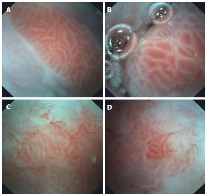
Figure 3 Barrett’s esophagus images obtained with E.
G. Scan™. A and B: Barrett’s without dysplasia; C and D: Barrett’s esophagus with low-grade dysplasia.
- Citation: Aedo MR, Zavala-González M&, Meixueiro-Daza A, Remes-Troche JM. Accuracy of transnasal endoscopy with a disposable esophagoscope compared to conventional endoscopy. World J Gastrointest Endosc 2014; 6(4): 128-136
- URL: https://www.wjgnet.com/1948-5190/full/v6/i4/128.htm
- DOI: https://dx.doi.org/10.4253/wjge.v6.i4.128


