Copyright
©2014 Baishideng Publishing Group Co.
World J Gastrointest Endosc. Mar 16, 2014; 6(3): 99-100
Published online Mar 16, 2014. doi: 10.4253/wjge.v6.i3.99
Published online Mar 16, 2014. doi: 10.4253/wjge.v6.i3.99
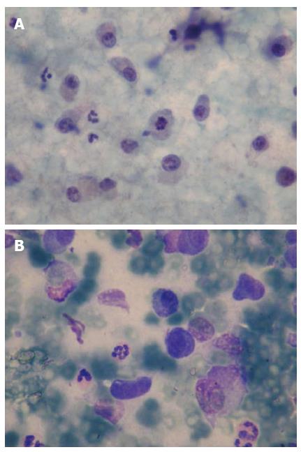
Figure 2 Cytopathologic findings of pancreatic mass obtained by endosonographic fine needle aspiration.
A: Neoplasic plasmacytoid cells, mitose in the middle (× 100, Papa-nikolaou stain); B: Neoplasic plasmacytoid cells (× 100, May-Grunwald-Giemsa).
- Citation: Akyuz F, Şahin D, Akyuz U, Vatansever S. Rare pancreas tumor mimicking adenocarcinoma: Extramedullary plasmacytoma. World J Gastrointest Endosc 2014; 6(3): 99-100
- URL: https://www.wjgnet.com/1948-5190/full/v6/i3/99.htm
- DOI: https://dx.doi.org/10.4253/wjge.v6.i3.99


