Copyright
©2014 Baishideng Publishing Group Inc.
World J Gastrointest Endosc. Dec 16, 2014; 6(12): 600-605
Published online Dec 16, 2014. doi: 10.4253/wjge.v6.i12.600
Published online Dec 16, 2014. doi: 10.4253/wjge.v6.i12.600
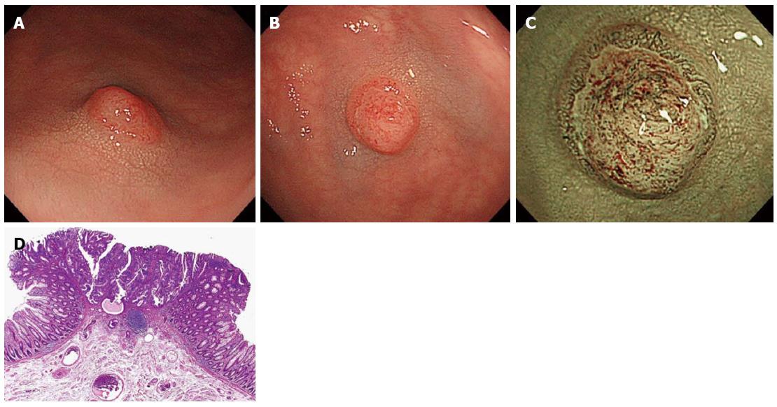
Figure 2 Invasive cancer 1 (S/C, 4 mm, IIa + IIc, Depressed type with NICE type 3).
A and B: 0-IIa+IIc lesion was shown in sigmoid colon; C: NBI-magnifying endoscopy showed the feature classified as NICE type 3; D: Pathological diagnosis was well differentiated tubular adenocarcinoma, pSM-M, ly(+), v(-), budding grade 0-1.
- Citation: Hattori S, Iwatate M, Sano W, Hasuike N, Kosaka H, Ikumoto T, Kotaka M, Ichiyanagi A, Ebisutani C, Hisano Y, Fujimori T, Sano Y. Narrow-band imaging observation of colorectal lesions using NICE classification to avoid discarding significant lesions. World J Gastrointest Endosc 2014; 6(12): 600-605
- URL: https://www.wjgnet.com/1948-5190/full/v6/i12/600.htm
- DOI: https://dx.doi.org/10.4253/wjge.v6.i12.600


