Copyright
©2014 Baishideng Publishing Group Inc.
World J Gastrointest Endosc. Dec 16, 2014; 6(12): 592-599
Published online Dec 16, 2014. doi: 10.4253/wjge.v6.i12.592
Published online Dec 16, 2014. doi: 10.4253/wjge.v6.i12.592
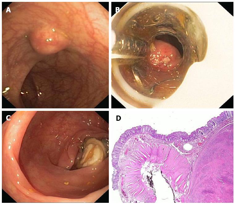
Figure 3 Endoscopic full thickness resection with the FTRD.
A: A 75 years old woman presented with a 1.5 cm subepithelial tumor in the descending colon; B: Endoscopic view with the FTRD mounted on a standard colonoscope; C: Resection site after endoscopic full thickness resection. The over-the-scope clips secures colonic wall patency; D: Histologic image (HE-staining) of the resection specimen showing one lateral resection margin. Note the cross-sectional view of the whole colonic wall on the left side. The tumor (leiomyoma) is shown on the right.
- Citation: Schmidt A, Bauder M, Riecken B, Caca K. Endoscopic resection of subepithelial tumors. World J Gastrointest Endosc 2014; 6(12): 592-599
- URL: https://www.wjgnet.com/1948-5190/full/v6/i12/592.htm
- DOI: https://dx.doi.org/10.4253/wjge.v6.i12.592


