Copyright
©2013 Baishideng Publishing Group Co.
World J Gastrointest Endosc. Aug 16, 2013; 5(8): 407-411
Published online Aug 16, 2013. doi: 10.4253/wjge.v5.i8.407
Published online Aug 16, 2013. doi: 10.4253/wjge.v5.i8.407
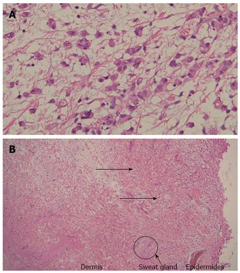
Figure 4 Pathological findings of the nodular lesion and the umbilical region.
A: Diffuse infiltration of oval or polygon cells with acidophilic cytoplasm was observed [hematoxylin and eosin (HE), × 200]; B: Histopathology of the umbilical region showed the invasion of tumor cells into the dermis (arrows) (HE, × 40).
- Citation: Tsuruya K, Matsushima M, Nakajima T, Fujisawa M, Shirakura K, Igarashi M, Koike J, Suzuki T, Mine T. Malignant peritoneal mesothelioma presenting umbilical hernia and Sister Mary Joseph’s nodule. World J Gastrointest Endosc 2013; 5(8): 407-411
- URL: https://www.wjgnet.com/1948-5190/full/v5/i8/407.htm
- DOI: https://dx.doi.org/10.4253/wjge.v5.i8.407


