Copyright
©2013 Baishideng Publishing Group Co.
World J Gastrointest Endosc. Jul 16, 2013; 5(7): 352-355
Published online Jul 16, 2013. doi: 10.4253/wjge.v5.i7.352
Published online Jul 16, 2013. doi: 10.4253/wjge.v5.i7.352
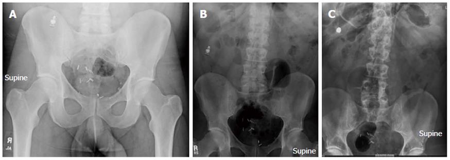
Figure 1 Abdominal X rays done at several time intervals following video capsule endoscopy showing retention of video capsule.
Several surgical clips are also present at pelvis. A: Follow up in 1 wk showing capsule in right lower quadrant of abdomen; B: Follow up in two months showing capsule in right mid abdomen laterally; C: Follow up after 4 years showing capsule in right upper quadrant of abdomen.
- Citation: Bhattarai M, Bansal P, Khan Y. Longest duration of retention of video capsule: A case report and literature review. World J Gastrointest Endosc 2013; 5(7): 352-355
- URL: https://www.wjgnet.com/1948-5190/full/v5/i7/352.htm
- DOI: https://dx.doi.org/10.4253/wjge.v5.i7.352


