Copyright
©2013 Baishideng Publishing Group Co.
World J Gastrointest Endosc. May 16, 2013; 5(5): 265-269
Published online May 16, 2013. doi: 10.4253/wjge.v5.i5.265
Published online May 16, 2013. doi: 10.4253/wjge.v5.i5.265
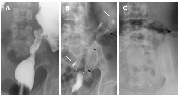
Figure 3 Radiographic images.
A: Stricture of the distal sigmoid colon outlined by luminal filling with barium using a rectal catheter (supine position); B: Biodegradable stent in situ immediately after insertion (supine position). The stent is radiolucent, but the radiopaque markers at the midpoint and at the ends are visible (black arrows). The margins of the stricture are marked with lipiodol (white arrows); C: Disappearance of radiopaque markers at 4 mo confirming complete stent degradation (erect position).
- Citation: Rodrigues C, Oliveira A, Santos L, Pires E, Deus J. Biodegradable stent for the treatment of a colonic stricture in Crohn’s disease. World J Gastrointest Endosc 2013; 5(5): 265-269
- URL: https://www.wjgnet.com/1948-5190/full/v5/i5/265.htm
- DOI: https://dx.doi.org/10.4253/wjge.v5.i5.265


