Copyright
©2013 Baishideng Publishing Group Co.
World J Gastrointest Endosc. Apr 16, 2013; 5(4): 174-179
Published online Apr 16, 2013. doi: 10.4253/wjge.v5.i4.174
Published online Apr 16, 2013. doi: 10.4253/wjge.v5.i4.174
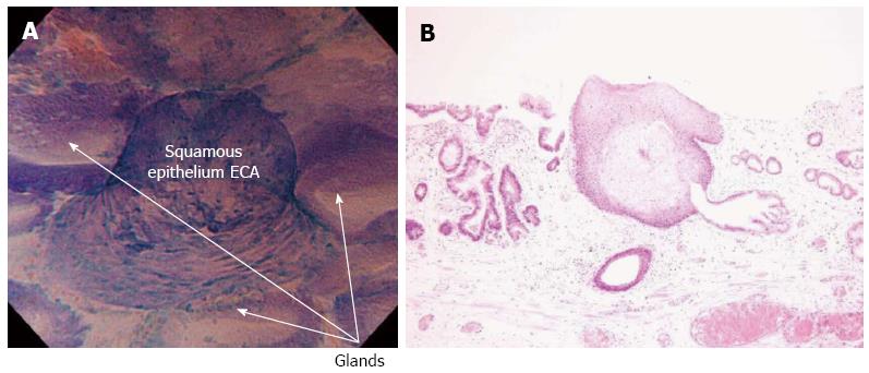
Figure 2 Endocytoscopy examination of histologically confirmed squamous cell islands within Barrett’s esophagus.
A: Endocytoscopy (ECS) examination shows squamous cell islands, within regular glandular structures of Barrett’s esophagus. According to ECS examination, squamous papillary structure is classified as ECA2, (round-shaped cells with different-sized small nuclei, suggestive of inflammatory changes); B: Histological examination (hematoxylin and eosin stain magnification) of biopsies from the same area as in Figure (A) confirmed the presence of a squamous papillary structure surrounded by Barrett’s glandular epithelium.
- Citation: Eleftheriadis N, Inoue H, Ikeda H, Onimaru M, Yoshida A, Hosoya T, Maselli R, Kudo SE. Endocytoscopic visualization of squamous cell islands within Barrett’s epithelium. World J Gastrointest Endosc 2013; 5(4): 174-179
- URL: https://www.wjgnet.com/1948-5190/full/v5/i4/174.htm
- DOI: https://dx.doi.org/10.4253/wjge.v5.i4.174


