Copyright
©2013 Baishideng Publishing Group Co.
World J Gastrointest Endosc. Mar 16, 2013; 5(3): 128-131
Published online Mar 16, 2013. doi: 10.4253/wjge.v5.i3.128
Published online Mar 16, 2013. doi: 10.4253/wjge.v5.i3.128
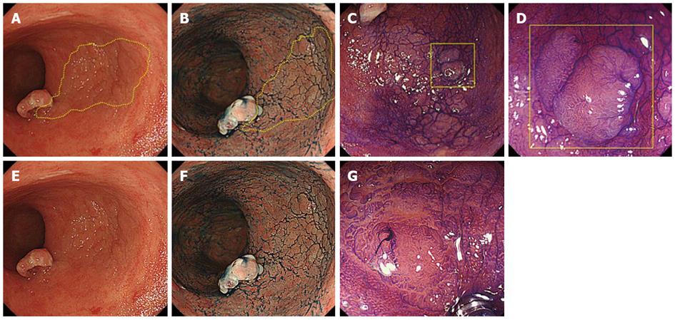
Figure 2 A flat adenoma, 35 mm in size, in the anterior wall of the low rectum.
A: The lesion was detected as a slight decline in vascular permeability on routine observation; B: Although we could highlight the irregular surface by spraying indigo carmine solution, it was difficult to trace the margin of the lesion; C, D: The surface structure of the lesion is composed mainly of IIIL pits with partially mixed IV pits on magnifying endoscopy with crystal violet staining; E: Figure A showing lesion borders without the aid of yellow lines; F: Figure B showing lesion borders without the aid of yellow lines; G: Magnifying endoscopy with crystal violet staining effectively delineates the margin of the lesion.
- Citation: Asayama N, Ikehara H, Yano H, Saito Y. Endoscopic submucosal dissection of multiple flat adenomas in the radiated rectum. World J Gastrointest Endosc 2013; 5(3): 128-131
- URL: https://www.wjgnet.com/1948-5190/full/v5/i3/128.htm
- DOI: https://dx.doi.org/10.4253/wjge.v5.i3.128


