Copyright
©2013 Baishideng Publishing Group Co.
World J Gastrointest Endosc. Mar 16, 2013; 5(3): 128-131
Published online Mar 16, 2013. doi: 10.4253/wjge.v5.i3.128
Published online Mar 16, 2013. doi: 10.4253/wjge.v5.i3.128
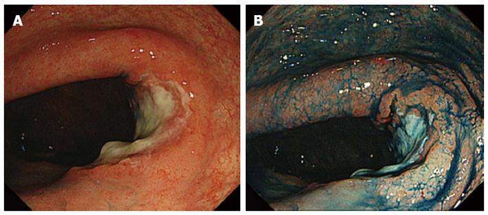
Figure 1 An advanced neoplasm, 35 mm in size, in the rectum.
A: Colonoscopy shows an ulcerative lesion in the rectum, the biopsy of which proved to be well differentiated adenocarcinoma; B: Chromoendoscopy with indigo carmine dye spray shows the lesion clearly.
- Citation: Asayama N, Ikehara H, Yano H, Saito Y. Endoscopic submucosal dissection of multiple flat adenomas in the radiated rectum. World J Gastrointest Endosc 2013; 5(3): 128-131
- URL: https://www.wjgnet.com/1948-5190/full/v5/i3/128.htm
- DOI: https://dx.doi.org/10.4253/wjge.v5.i3.128


