Copyright
©2013 Baishideng Publishing Group Co.
World J Gastrointest Endosc. Dec 16, 2013; 5(12): 605-609
Published online Dec 16, 2013. doi: 10.4253/wjge.v5.i12.605
Published online Dec 16, 2013. doi: 10.4253/wjge.v5.i12.605
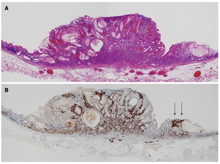
Figure 3 Histological examination of the resected specimen.
A: Microscopic examination of the completely resected specimen revealed a neuroendocrine tumor presenting in both the mucosal layer and submucosal layer (hematoxylin and eosin staining); B: Immunohistochemistry for synaptophysin showed that the tumor extended through the normal gland ducts randomly. Enterochromaffin-like cell micro-nests were observed below the normal mucosa (arrow).
- Citation: Hirai M, Matsumoto K, Ueyama H, Fukushima H, Murakami T, Sasaki H, Nagahara A, Yao T, Watanabe S. A case of neuroendocrine tumor G1 with unique histopathological growth progress. World J Gastrointest Endosc 2013; 5(12): 605-609
- URL: https://www.wjgnet.com/1948-5190/full/v5/i12/605.htm
- DOI: https://dx.doi.org/10.4253/wjge.v5.i12.605


