Copyright
©2013 Baishideng Publishing Group Co.
World J Gastrointest Endosc. Nov 16, 2013; 5(11): 581-583
Published online Nov 16, 2013. doi: 10.4253/wjge.v5.i11.581
Published online Nov 16, 2013. doi: 10.4253/wjge.v5.i11.581
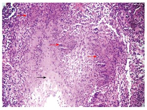
Figure 3 Histopathological examination of esophageal ulcer biopsy showing epitheloid cell granulomas (red arrows) with caseation (black arrow) in the exudate suggestive of esophageal tuberculosis (HE stain x 10).
- Citation: Jain SS, Somani PO, Mahey RC, Shah DK, Contractor QQ, Rathi PM. Esophageal tuberculosis presenting with hematemesis. World J Gastrointest Endosc 2013; 5(11): 581-583
- URL: https://www.wjgnet.com/1948-5190/full/v5/i11/581.htm
- DOI: https://dx.doi.org/10.4253/wjge.v5.i11.581


