Copyright
©2013 Baishideng Publishing Group Co.
World J Gastrointest Endosc. Oct 16, 2013; 5(10): 514-518
Published online Oct 16, 2013. doi: 10.4253/wjge.v5.i10.514
Published online Oct 16, 2013. doi: 10.4253/wjge.v5.i10.514
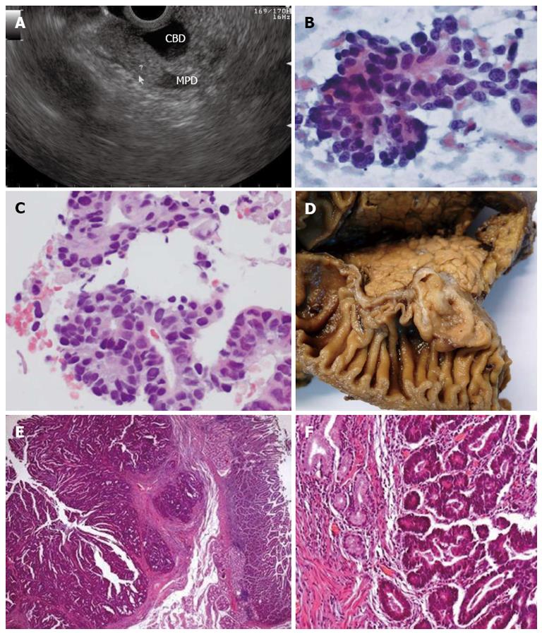
Figure 2 Endoscopic ultrasonography and pathological findings of an ampullary carcinoma.
A: Mildly hyperechoic third-layer lesion adjacent to the ampulla, compressing the bile duct; B, C: Fine-needle aspiration, × 400 magnification; B: Smears with acinar groups, irregularly distributed nuclei, coarse chromatin, conspicuous nucleoli (Papanicolaou); C: Cell-block preparation of aspirated sample [hematoxylin and eosin (HE)]; D: Surgical pathology specimen confirming the full excision of an ampullary carcinoma; E: Ampullary area well-differentiated adenocarcinoma, HE × 25; F: HE × 100.
- Citation: Figueiredo PC, Pinto-Marques P, Mendonça E, Oliveira P, Brito M, Serra D. Duodenal subepithelial hyperechoic lesions of the third layer: Not always a lipoma. World J Gastrointest Endosc 2013; 5(10): 514-518
- URL: https://www.wjgnet.com/1948-5190/full/v5/i10/514.htm
- DOI: https://dx.doi.org/10.4253/wjge.v5.i10.514


