Copyright
©2012 Baishideng.
World J Gastrointest Endosc. Sep 16, 2012; 4(9): 421-428
Published online Sep 16, 2012. doi: 10.4253/wjge.v4.i9.421
Published online Sep 16, 2012. doi: 10.4253/wjge.v4.i9.421
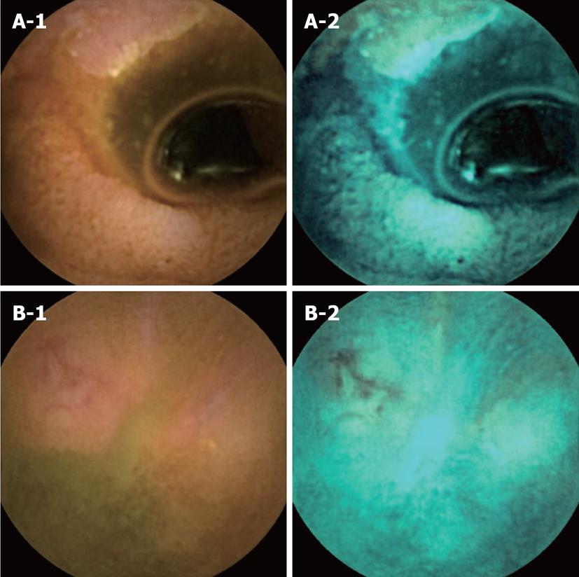
Figure 3 Two cases of small bowel lesions were missed with flexible spectral color enhancement imaging.
A-1: Conventional capsule endoscopy (CE) image; A-2: FICE set 2. An ulcer was missed with flexible spectral color enhancement (FICE) imaging and only detected with conventional CE imaging; B-1: Conventional CE image; B-2: FICE set 2. An ulcer was missed with FICE imaging and only detected with conventional CE imaging.
- Citation: Matsumura T, Arai M, Sato T, Nakagawa T, Maruoka D, Tsuboi M, Hata S, Arai E, Katsuno T, Imazeki F, Yokosuka O. Efficacy of computed image modification of capsule endoscopy in patients with obscure gastrointestinal bleeding. World J Gastrointest Endosc 2012; 4(9): 421-428
- URL: https://www.wjgnet.com/1948-5190/full/v4/i9/421.htm
- DOI: https://dx.doi.org/10.4253/wjge.v4.i9.421


