Copyright
©2012 Baishideng.
World J Gastrointest Endosc. Sep 16, 2012; 4(9): 387-397
Published online Sep 16, 2012. doi: 10.4253/wjge.v4.i9.387
Published online Sep 16, 2012. doi: 10.4253/wjge.v4.i9.387
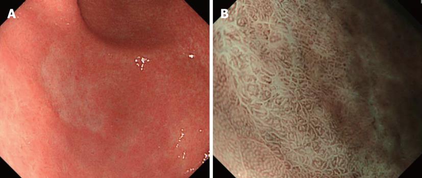
Figure 13 A case of gastric undifferentiated adenocarcinoma.
A: A pale concave lesion was noted in the antrum on the anterior wall side of the greater curvature (undifferentiated cancer was diagnosed by biopsy); B: Findings on narrow band imaging-combined magnifying observation of A: Only swollen intervening parts and increased intervals between the white lines were observed, and there were no abnormal blood vessels. The gland structures in the surrounding normal mucosa appeared different from those of the concave surface.
- Citation: Nonaka K, Nishimura M, Kita H. Role of narrow band imaging in endoscopic submucosal dissection. World J Gastrointest Endosc 2012; 4(9): 387-397
- URL: https://www.wjgnet.com/1948-5190/full/v4/i9/387.htm
- DOI: https://dx.doi.org/10.4253/wjge.v4.i9.387


