Copyright
©2012 Baishideng.
World J Gastrointest Endosc. Sep 16, 2012; 4(9): 387-397
Published online Sep 16, 2012. doi: 10.4253/wjge.v4.i9.387
Published online Sep 16, 2012. doi: 10.4253/wjge.v4.i9.387
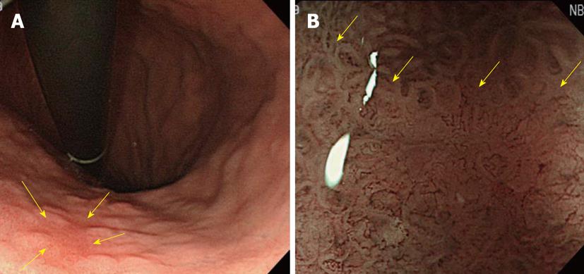
Figure 12 A case of small flat-type differentiated early-stage gastric cancer with an unclear boundary.
A: A small flat-type differentiated early-stage gastric cancer of about 6 mm with an unclear boundary was present in the small curvature of the lower body of the stomach (arrows), but visualization of the lesion by conventional observation was difficult; B: Findings on narrow band imaging-combined magnifying observation: The border between the tumor and non-tumorous region was clearly observed, and apparently abnormal blood vessels were present inside the demarcation line (arrows).
- Citation: Nonaka K, Nishimura M, Kita H. Role of narrow band imaging in endoscopic submucosal dissection. World J Gastrointest Endosc 2012; 4(9): 387-397
- URL: https://www.wjgnet.com/1948-5190/full/v4/i9/387.htm
- DOI: https://dx.doi.org/10.4253/wjge.v4.i9.387


