Copyright
©2012 Baishideng.
World J Gastrointest Endosc. Sep 16, 2012; 4(9): 387-397
Published online Sep 16, 2012. doi: 10.4253/wjge.v4.i9.387
Published online Sep 16, 2012. doi: 10.4253/wjge.v4.i9.387
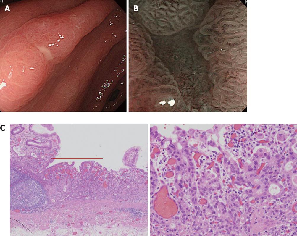
Figure 11 Differentiation of benignity and malignancy of a small depressed lesion.
A: A solitary erosion was present in the antrum on the small curvature side. Differentiation of benignity and malignancy by conventional observation was difficult; B: The gland structure on the erosion surface was apparently different from the surrounding region, and a few abnormal micro blood vessels with an abnormal distribution and irregular width were present in this region; C: The lesion was an intramucosal moderately differentiated adenocarcinoma on pathological examination after endoscopic submucosal dissection. HE, original magnification, × 100 (left), original magnification, × 400 (right).
- Citation: Nonaka K, Nishimura M, Kita H. Role of narrow band imaging in endoscopic submucosal dissection. World J Gastrointest Endosc 2012; 4(9): 387-397
- URL: https://www.wjgnet.com/1948-5190/full/v4/i9/387.htm
- DOI: https://dx.doi.org/10.4253/wjge.v4.i9.387


