Copyright
©2012 Baishideng.
World J Gastrointest Endosc. Sep 16, 2012; 4(9): 387-397
Published online Sep 16, 2012. doi: 10.4253/wjge.v4.i9.387
Published online Sep 16, 2012. doi: 10.4253/wjge.v4.i9.387
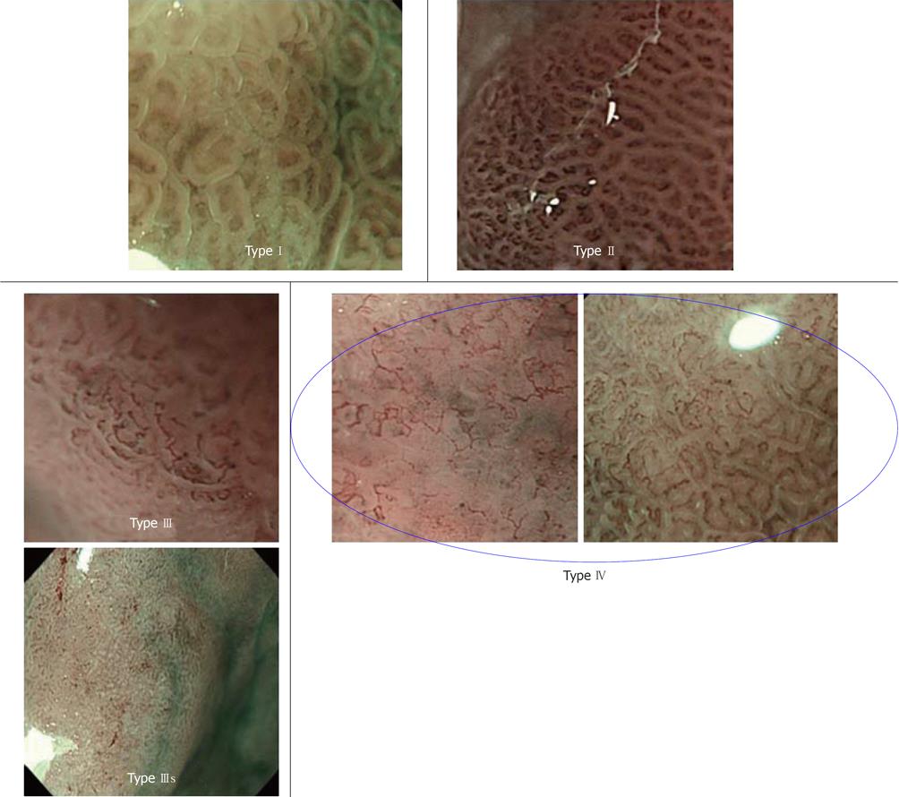
Figure 10 Narrow band imaging typing.
Type I: “Clear” mucosal microstructure and “unclear” microvessel image; Type II: “Clear” mucosal microstructure and “clear” microvessel image; Type III: “Clear” mucosal microstructure and “abnormal” microvessel image; Type IIIs: Very dense arrangement of small glands and “dots” or “unclear” microvessel image; Type IV: “Obscured” mucosal microstructure and “abnormal” microvessel image.
- Citation: Nonaka K, Nishimura M, Kita H. Role of narrow band imaging in endoscopic submucosal dissection. World J Gastrointest Endosc 2012; 4(9): 387-397
- URL: https://www.wjgnet.com/1948-5190/full/v4/i9/387.htm
- DOI: https://dx.doi.org/10.4253/wjge.v4.i9.387


