Copyright
©2012 Baishideng.
World J Gastrointest Endosc. Aug 16, 2012; 4(8): 373-375
Published online Aug 16, 2012. doi: 10.4253/wjge.v4.i8.373
Published online Aug 16, 2012. doi: 10.4253/wjge.v4.i8.373
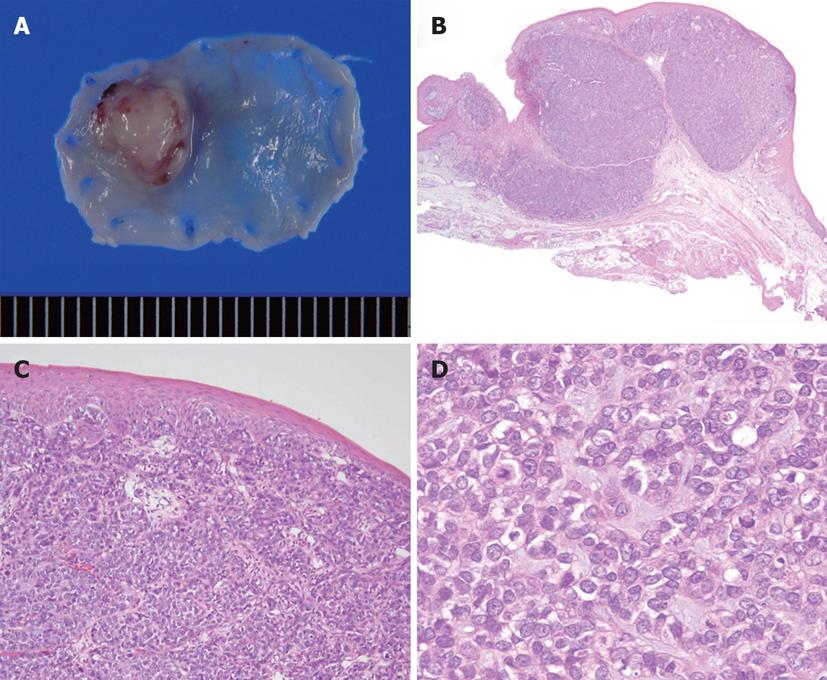
Figure 2 Resected specimen.
A: In a fresh resected specimen, the lesion is defined as 0-I and measures 7 mm × 6 mm; B: The histological diagnosis is basaloid squamous carcinoma (BSC), and tumor invasion depth is limited to the muscularis mucosae (HE, × 40); C: BSC is located in the lamina propria mucosae, and covered by normal squamous epithelium (HE, × 100); D: The tumor consists of oval cells like basal cells (HE, × 200).
- Citation: Nakamura M, Nishikawa J, Suenaga S, Okamoto T, Okamoto F, Miura O, Sakaida I. A case of EMRC for basaloid squamous carcinoma of the cervical esophagus. World J Gastrointest Endosc 2012; 4(8): 373-375
- URL: https://www.wjgnet.com/1948-5190/full/v4/i8/373.htm
- DOI: https://dx.doi.org/10.4253/wjge.v4.i8.373


