Copyright
©2012 Baishideng.
World J Gastrointest Endosc. Aug 16, 2012; 4(8): 373-375
Published online Aug 16, 2012. doi: 10.4253/wjge.v4.i8.373
Published online Aug 16, 2012. doi: 10.4253/wjge.v4.i8.373
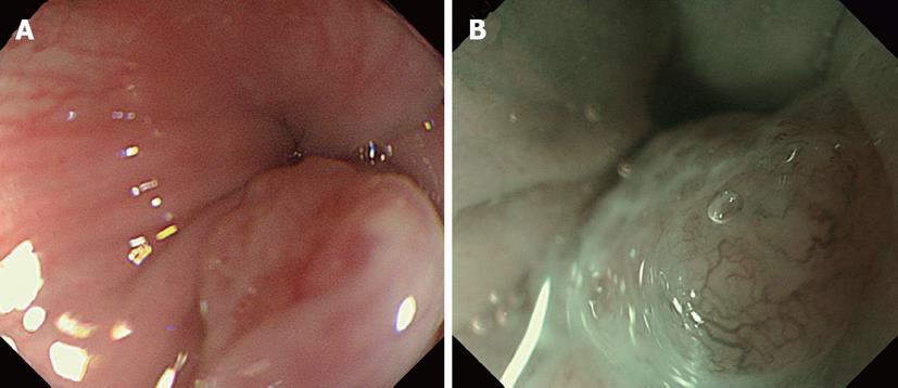
Figure 1 Endoscopic images.
A: An endoscopic image shows a protruding lesion located in the cervical esophagus; B: Magnifying endoscopy with narrow-band imaging shows microvessels of type 4 M of the Arima classification.
- Citation: Nakamura M, Nishikawa J, Suenaga S, Okamoto T, Okamoto F, Miura O, Sakaida I. A case of EMRC for basaloid squamous carcinoma of the cervical esophagus. World J Gastrointest Endosc 2012; 4(8): 373-375
- URL: https://www.wjgnet.com/1948-5190/full/v4/i8/373.htm
- DOI: https://dx.doi.org/10.4253/wjge.v4.i8.373


