Copyright
©2012 Baishideng.
World J Gastrointest Endosc. Aug 16, 2012; 4(8): 356-361
Published online Aug 16, 2012. doi: 10.4253/wjge.v4.i8.356
Published online Aug 16, 2012. doi: 10.4253/wjge.v4.i8.356
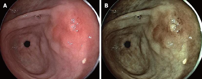
Figure 5 In some cases, a partially reddish change is accompanied on the tumor surface similar to depressed type cancer.
A: Conventional image (EG590-ZW) in a distant view reveals an elevated lesion with slightly reddish portion in the posterior wall of antrum; B: Flexible spectral imaging color enhancement image enhances a reddish portion on tumor surface with more contrasting demarcation line. In addition, tumor margin of flat area in the right side of this figure can be more clearly visualized than conventional image.
- Citation: Osawa H, Yamamoto H, Miura Y, Yoshizawa M, Sunada K, Satoh K, Sugano K. Diagnosis of extent of early gastric cancer using flexible spectral imaging color enhancement. World J Gastrointest Endosc 2012; 4(8): 356-361
- URL: https://www.wjgnet.com/1948-5190/full/v4/i8/356.htm
- DOI: https://dx.doi.org/10.4253/wjge.v4.i8.356


