Copyright
©2012 Baishideng.
World J Gastrointest Endosc. Aug 16, 2012; 4(8): 356-361
Published online Aug 16, 2012. doi: 10.4253/wjge.v4.i8.356
Published online Aug 16, 2012. doi: 10.4253/wjge.v4.i8.356
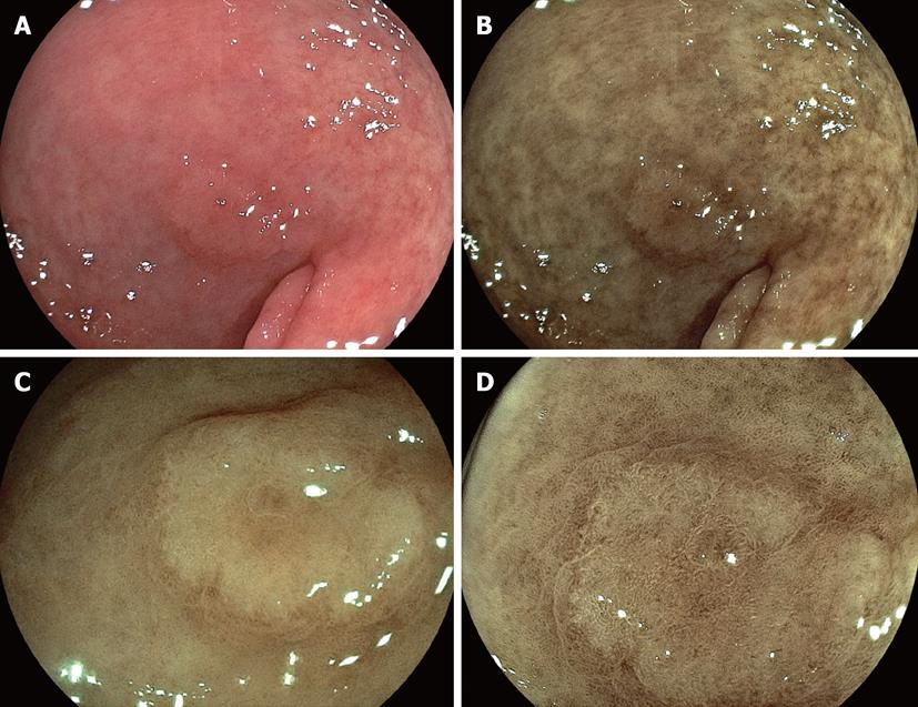
Figure 2 Some depressed cancers are shown as whitish lesion by conventional endoscopy.
A: Conventional image (EG590-ZW) reveals a slightly whitish mucosal change in the anterior wall of antrum; B: Flexible spectral imaging color enhancement (FICE) image enhances a whitish cancerous lesion and can determine a line of demarcation between cancer and surrounding mucosa; C: FICE image in a close-up view (EG590-WR) detects with precision a clearer demarcation line; D: FICE image with half magnification (EG590-ZW) reveals a finer microstructural pattern on mucosal surface of cancer and higher contrasting mucosa between cancer and the surrounding area, leading a clearer demarcation line.
- Citation: Osawa H, Yamamoto H, Miura Y, Yoshizawa M, Sunada K, Satoh K, Sugano K. Diagnosis of extent of early gastric cancer using flexible spectral imaging color enhancement. World J Gastrointest Endosc 2012; 4(8): 356-361
- URL: https://www.wjgnet.com/1948-5190/full/v4/i8/356.htm
- DOI: https://dx.doi.org/10.4253/wjge.v4.i8.356


