Copyright
©2012 Baishideng Publishing Group Co.
World J Gastrointest Endosc. Jul 16, 2012; 4(7): 335-338
Published online Jul 16, 2012. doi: 10.4253/wjge.v4.i7.335
Published online Jul 16, 2012. doi: 10.4253/wjge.v4.i7.335
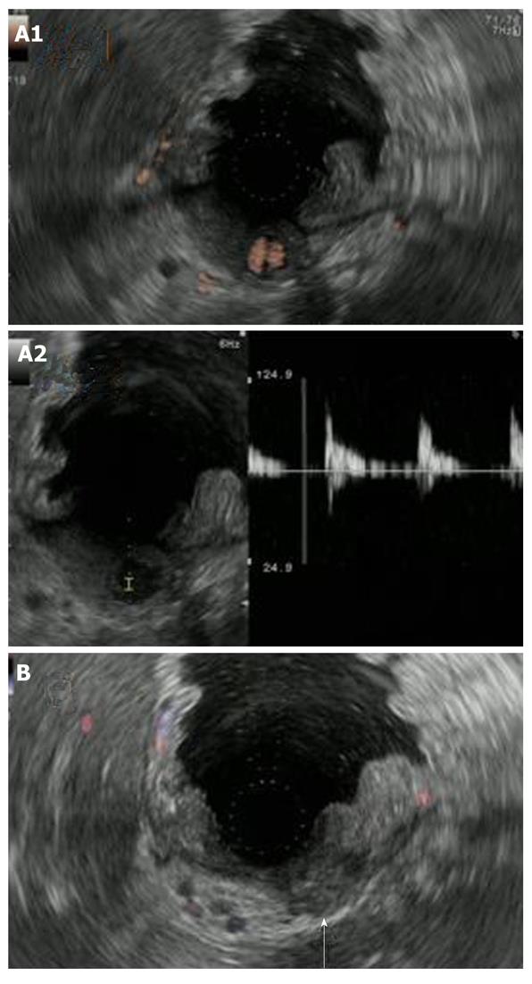
Figure 4 Endoscopic ultrasonography findings.
A1: Endoscopic ultrasonography showed an anechoic region whose entire periphery was hypoechoic beneath the gastric mucosa. Power Doppler showed blood flow in the anechoic region. Upper gastrointestinal endoscopy showed a 2 cm, submucosal tumor-like protrusion with a red, eroded upper region located in the lesser curvature of middle of the body of the stomach (arrow); A2: Pulsed wave Doppler showed pulsatile blood flow in the anechoic region. This finding led to the diagnosis of an aneurysm; B: The cessation of blood flow to the pseudoaneurysm was confirmed with endoscopic ultrasonography which was performed 1 wk after treatment (arrow).
- Citation: Fukatsu K, Ueda K, Maeda H, Yamashita Y, Itonaga M, Mori Y, Moribata K, Shingaki N, Deguchi H, Enomoto S, Inoue I, Maekita T, Iguchi M, Tamai H, Kato J, Ichinose M. A case of chronic pancreatitis in which endoscopic ultrasonography was effective in the diagnosis of a pseudoaneurysm. World J Gastrointest Endosc 2012; 4(7): 335-338
- URL: https://www.wjgnet.com/1948-5190/full/v4/i7/335.htm
- DOI: https://dx.doi.org/10.4253/wjge.v4.i7.335


