Copyright
©2012 Baishideng Publishing Group Co.
World J Gastrointest Endosc. Jul 16, 2012; 4(7): 331-334
Published online Jul 16, 2012. doi: 10.4253/wjge.v4.i7.331
Published online Jul 16, 2012. doi: 10.4253/wjge.v4.i7.331
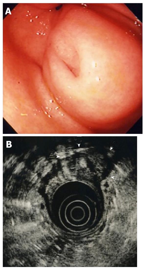
Figure 1 Esophagogastroduodenoscopic and endoscopic ultrasonographic images of submucosal lesion at the first examination.
A: Esophagogastroduodenoscopy. Submucosal lesion was visible in the antrum of the stomach; B: Endoscopic ultrasonography. Lesion (arrow heads) located in the third layer (submucosa) with a diameter of 20 mm, slightly hypoechoic internal echo, and anechoic lumen, regarded as the duct.
- Citation: Watanabe K, Irisawa A, Hikichi T, Takagi T, Shibukawa G, Sato M, Obara K, Ohira H. Acute inflammation occurring in gastric aberrant pancreas followed up by endoscopic ultrasonography. World J Gastrointest Endosc 2012; 4(7): 331-334
- URL: https://www.wjgnet.com/1948-5190/full/v4/i7/331.htm
- DOI: https://dx.doi.org/10.4253/wjge.v4.i7.331


