Copyright
©2012 Baishideng Publishing Group Co.
World J Gastrointest Endosc. Jun 16, 2012; 4(6): 247-259
Published online Jun 16, 2012. doi: 10.4253/wjge.v4.i6.247
Published online Jun 16, 2012. doi: 10.4253/wjge.v4.i6.247
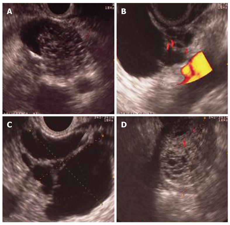
Figure 1 Serous cystoadenoma.
A: Microcystic area, centrally located; B: Beside microcystic area; C: Peripheral and internal septa microcystic area, lobulate contour; D: Pseudosolid form.
- Citation: Barresi L, Tarantino I, Granata A, Curcio G, Traina M. Pancreatic cystic lesions: How endoscopic ultrasound morphology and endoscopic ultrasound fine needle aspiration help unlock the diagnostic puzzle. World J Gastrointest Endosc 2012; 4(6): 247-259
- URL: https://www.wjgnet.com/1948-5190/full/v4/i6/247.htm
- DOI: https://dx.doi.org/10.4253/wjge.v4.i6.247


