Copyright
©2012 Baishideng Publishing Group Co.
World J Gastrointest Endosc. Jun 16, 2012; 4(6): 218-226
Published online Jun 16, 2012. doi: 10.4253/wjge.v4.i6.218
Published online Jun 16, 2012. doi: 10.4253/wjge.v4.i6.218
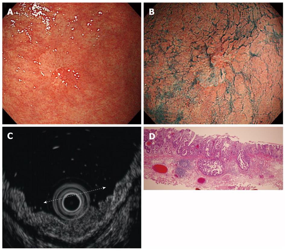
Figure 14 Findings for an sm1 cancer of the stomach.
A: Endoscopic features. A white, flat lesion with central elevation was located on the greater curvature of the angle; B: Endoscopic features after indigo carmine dye. Biopsy specimens showed adenocarcinoma; C: Endoscopic ultrasound (EUS) features. The white dotted line indicates the extent of the lesion. EUS revealed a thickness of the second layer and a slightly irregular third layer; D: Pathological findings. The tumor was invading the submucosal layer to a 400 μm depth. (Hematoxylin and eosin stain, × 40).
- Citation: Yoshinaga S, Oda I, Nonaka S, Kushima R, Saito Y. Endoscopic ultrasound using ultrasound probes for the diagnosis of early esophageal and gastric cancers. World J Gastrointest Endosc 2012; 4(6): 218-226
- URL: https://www.wjgnet.com/1948-5190/full/v4/i6/218.htm
- DOI: https://dx.doi.org/10.4253/wjge.v4.i6.218


