Copyright
©2012 Baishideng Publishing Group Co.
World J Gastrointest Endosc. Mar 16, 2012; 4(3): 57-64
Published online Mar 16, 2012. doi: 10.4253/wjge.v4.i3.57
Published online Mar 16, 2012. doi: 10.4253/wjge.v4.i3.57
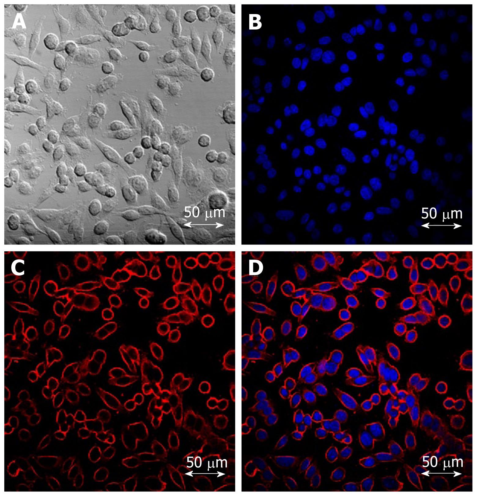
Figure 2 Confocal laser scanning microscopy images of colon cancer cells treated with cetuximab-conjugated magnetofluorescent nanoparticles.
A: A bright field image; B: Nuclear staining with DAPI; C: RITC (Rhodamine B isothiocyanate) fluorescent image; D: Overlay of B and C. Cellular uptake was so significant that the outer cell membranes of HCT116 cells can be clearly delineated by the images of the particles.
- Citation: Kwon YS, Cho YS, Yoon TJ, Kim HS, Choi MG. Recent advances in targeted endoscopic imaging: Early detection of gastrointestinal neoplasms. World J Gastrointest Endosc 2012; 4(3): 57-64
- URL: https://www.wjgnet.com/1948-5190/full/v4/i3/57.htm
- DOI: https://dx.doi.org/10.4253/wjge.v4.i3.57


