Copyright
©2012 Baishideng Publishing Group Co.
World J Gastrointest Endosc. Mar 16, 2012; 4(3): 50-56
Published online Mar 16, 2012. doi: 10.4253/wjge.v4.i3.50
Published online Mar 16, 2012. doi: 10.4253/wjge.v4.i3.50
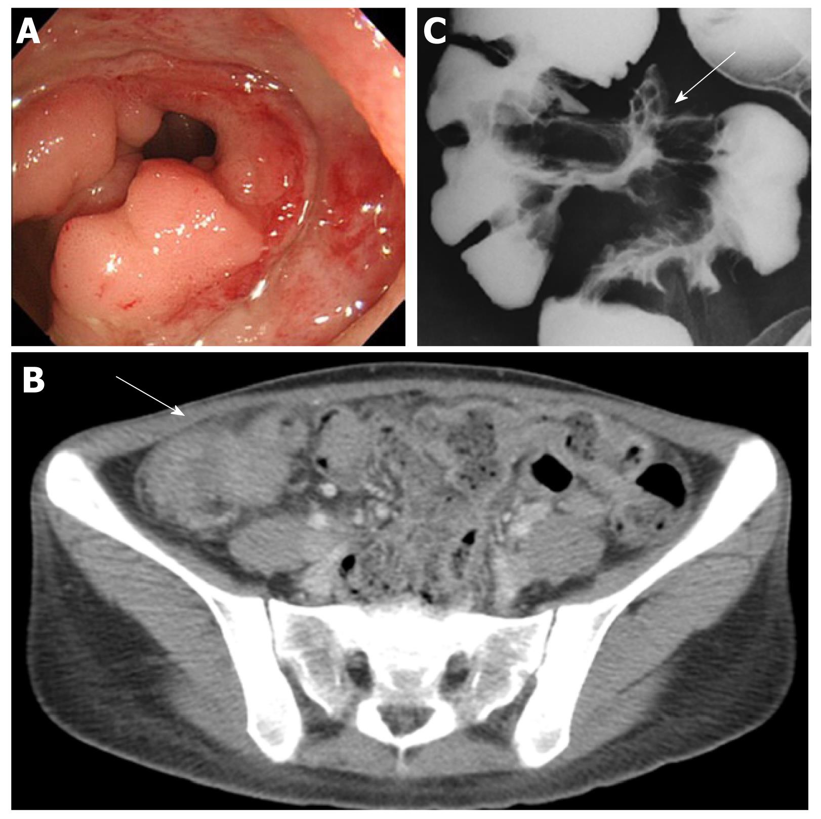
Figure 5 Behçet’s disease in a 25-year-old woman with abdominal pain and diarrhea.
A: Colonoscopy showed a large punched-out ulcer with elevated margins in the terminal ileum; B: Contrast-enhanced computed tomography scan of the abdomen showed a mass-like lesion with unevenly thickened bowel wall of the ileocecal region (arrow); C: Small bowel barium radiography disclosed the large ulcer (arrow) with convergence of mucosal folds in the terminal ileum.
- Citation: Hokama A, Kishimoto K, Ihama Y, Kobashigawa C, Nakamoto M, Hirata T, Kinjo N, Higa F, Tateyama M, Kinjo F, Iseki K, Kato S, Fujita J. Endoscopic and radiographic features of gastrointestinal involvement in vasculitis. World J Gastrointest Endosc 2012; 4(3): 50-56
- URL: https://www.wjgnet.com/1948-5190/full/v4/i3/50.htm
- DOI: https://dx.doi.org/10.4253/wjge.v4.i3.50


