Copyright
©2012 Baishideng Publishing Group Co.
World J Gastrointest Endosc. Dec 16, 2012; 4(12): 545-555
Published online Dec 16, 2012. doi: 10.4253/wjge.v4.i12.545
Published online Dec 16, 2012. doi: 10.4253/wjge.v4.i12.545
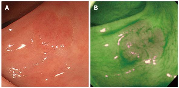
Figure 3 Autofluorescence imaging.
A: IIa polyp 14 mm in diameter (White-light endoscopy figure); B: In autofluorescence imaging, the normal mucosa was detected by green color and the neoplastic polyp was detected by magenta color.
- Citation: Yoshida N, Yagi N, Yanagisawa A, Naito Y. Image-enhanced endoscopy for diagnosis of colorectal tumors in view of endoscopic treatment. World J Gastrointest Endosc 2012; 4(12): 545-555
- URL: https://www.wjgnet.com/1948-5190/full/v4/i12/545.htm
- DOI: https://dx.doi.org/10.4253/wjge.v4.i12.545


