Copyright
©2012 Baishideng Publishing Group Co.
World J Gastrointest Endosc. Nov 16, 2012; 4(11): 489-499
Published online Nov 16, 2012. doi: 10.4253/wjge.v4.i11.489
Published online Nov 16, 2012. doi: 10.4253/wjge.v4.i11.489
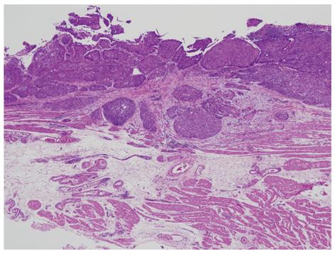
Figure 13 pT1a-superficial muscularis mucosae case.
The lower portion of the picture shows two-layered muscularis mucosae. In esophageal cancers with pT1a-MM (M3), the depth of invasion is divided into three (pT1a-superficial muscularis mucosae, pT1a-lamina propria mucosae and pT1a-deep muscularis mucosae).
- Citation: Nagata K, Shimizu M. Pathological evaluation of gastrointestinal endoscopic submucosal dissection materials based on Japanese guidelines. World J Gastrointest Endosc 2012; 4(11): 489-499
- URL: https://www.wjgnet.com/1948-5190/full/v4/i11/489.htm
- DOI: https://dx.doi.org/10.4253/wjge.v4.i11.489


