Copyright
©2012 Baishideng Publishing Group Co.
World J Gastrointest Endosc. Nov 16, 2012; 4(11): 489-499
Published online Nov 16, 2012. doi: 10.4253/wjge.v4.i11.489
Published online Nov 16, 2012. doi: 10.4253/wjge.v4.i11.489
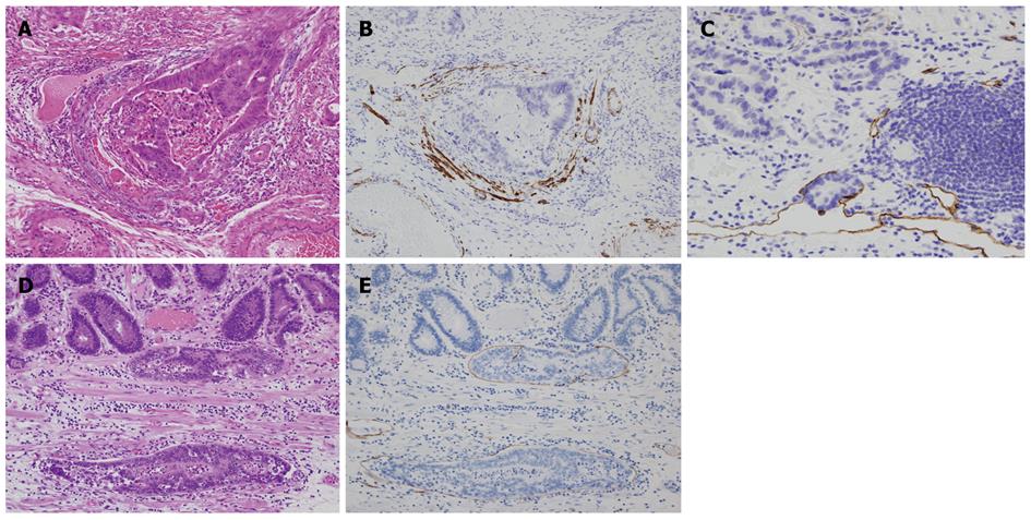
Figure 4 Evaluation of vessel invasion and lymphatic invasion.
Venous invasion is evaluated by using a double staining with Victoria blue and hematoxylin-eosin (A) and immunohistochemistry of desmin (B). Lymphatic invasion is demonstrated by immunostaining of D2-40 (C). Lymphatic invasion is noted in the lamina propria (D: Hematoxylin-eosin stain; E: Immunohistochemistry of D2-40).
- Citation: Nagata K, Shimizu M. Pathological evaluation of gastrointestinal endoscopic submucosal dissection materials based on Japanese guidelines. World J Gastrointest Endosc 2012; 4(11): 489-499
- URL: https://www.wjgnet.com/1948-5190/full/v4/i11/489.htm
- DOI: https://dx.doi.org/10.4253/wjge.v4.i11.489


