Copyright
©2011 Baishideng Publishing Group Co.
World J Gastrointest Endosc. Aug 16, 2011; 3(8): 157-161
Published online Aug 16, 2011. doi: 10.4253/wjge.v3.i8.157
Published online Aug 16, 2011. doi: 10.4253/wjge.v3.i8.157
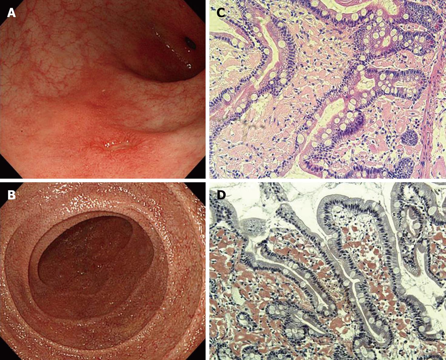
Figure 2 Endoscopic views of amyloid A amyloidosis in a 45-year-old woman with rheumatoid arthritis.
A: A round ulcer surrounded by longitudinal reddish mucosa is presented in the gastric antrum. Histopathological examination confirmed amyloid deposition; B: Fine granular mucosa in the descending duodenum; C: Biopsy of the duodenal lesion showing marked amorphous eosinophilic deposition in the lamina propria mucosae (HE, × 100); D: Congo red staining showing amyloid deposition (× 100).
- Citation: Hokama A, Kishimoto K, Nakamoto M, Kobashigawa C, Hirata T, Kinjo N, Kinjo F, Kato S, Fujita J. Endoscopic and histopathological features of gastrointestinal amyloidosis. World J Gastrointest Endosc 2011; 3(8): 157-161
- URL: https://www.wjgnet.com/1948-5190/full/v3/i8/157.htm
- DOI: https://dx.doi.org/10.4253/wjge.v3.i8.157


