Copyright
©2011 Baishideng Publishing Group Co.
World J Gastrointest Endosc. Jul 16, 2011; 3(7): 154-156
Published online Jul 16, 2011. doi: 10.4253/wjge.v3.i7.154
Published online Jul 16, 2011. doi: 10.4253/wjge.v3.i7.154
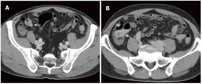
Figure 1 Contrast-enhanced computed tomography scans on admission.
A: In the arterial phase, a diverticulum of the terminal ileum was seen and leakage of the contrast medium into the ileal lumen around the diverticulum was also visualized (arrow); B: In the venous phase, the contrast medium spread to the ileocecal valve (arrow).
- Citation: Iwamuro M, Hanada M, Kominami Y, Higashi R, Mizuno M, Yamamoto K. Endoscopic hemostasis for hemorrhage from an ileal diverticulum. World J Gastrointest Endosc 2011; 3(7): 154-156
- URL: https://www.wjgnet.com/1948-5190/full/v3/i7/154.htm
- DOI: https://dx.doi.org/10.4253/wjge.v3.i7.154


