Copyright
©2011 Baishideng Publishing Group Co.
World J Gastrointest Endosc. Jun 16, 2011; 3(6): 124-128
Published online Jun 16, 2011. doi: 10.4253/wjge.v3.i6.124
Published online Jun 16, 2011. doi: 10.4253/wjge.v3.i6.124
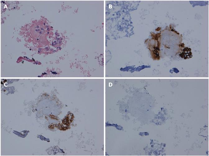
Figure 3 Omental nodule.
A: Hematoxylin and eosin immunohistochemitry of the omental nodule; B: Cytokeratin-19 immunohistochemistry of the omental nodule (high sensitivity, high specificity); C: Carcinogenic-embryonic antigen immunohistochemistry of the omental nodule (high sensitivity, low specificity); D: Wilm’s Tumor-1 immunohistochemistry of the omental nodule (mesenchymal marker).
- Citation: Rial NS, Gilchrist KB, Henderson JT, Bhattacharyya AK, Boyer TD, Nadir A, Cunningham JT. Endoscopic ultrasound with biopsy of omental mass for cholangiocarcinoma diagnosis in cirrhosis. World J Gastrointest Endosc 2011; 3(6): 124-128
- URL: https://www.wjgnet.com/1948-5190/full/v3/i6/124.htm
- DOI: https://dx.doi.org/10.4253/wjge.v3.i6.124


